Compact Bone Diagram Microscope - 1 - Magnification by a microscope is the product of the individual magnifying ability of the oculars and the objectives.
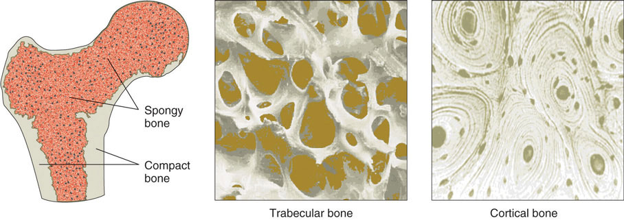
Compact Bone Diagram Microscope - 1 - Magnification by a microscope is the product of the individual magnifying ability of the oculars and the objectives.. Fruit microscopic structure of long bone. Sclerostin inhibits bone formation mostly by antagonizing lrp5/6, thus inhibiting wnt signaling. Electron microscope image of trabecular bone (x100 magnification). This human bone section shows the haversian canal (or osteon) structure of compact bone tissue. Compact bone, spongy if you looked at it through a microscope, however, you would see that it's actually filled with many very.
If you look at compact bone under the microscope, you will observe a highly organized arrangement of concentric circles that look like tree trunks. Electron microscope image of trabecular bone (x100 magnification). Spongy bone and compact bone. Trouvez des images de stock de compact bone micrograph light microscopy bone en hd et des millions d'autres photos, illustrations et images vectorielles de stock libres de droits dans la collection shutterstock. Histology of human tissue, show skin as seen under the microscope.
Ultrastructure Of Bone Components Structure Teachmeanatomy from teachmeanatomy.info Compact bone, also called cortical bone, is the hard, stiff, smooth, thin, white bone tissue that surrounds all bones in the human body. 2 compact bone we know that compact bone is very dense it is also very complex when viewed under a microscope. The ground substance of bone is arranged in concentrated layers (lamellae) round the small canals which run parallel to the long axis (shaft) of the bone. Compact bone, spongy if you looked at it through a microscope, however, you would see that it's actually filled with many very. Having been constructed in the 16th century, microscopes have revolutionalized science with their ability to magnify small objects such as microbial cells, producing images with definitive structures that are. Compact bone diagram bone cross section diagram file624 diagram of compact bone new. Confocal microscopy of fibroblast cells. To better understand the structure and function of a microscope, we need to take a look at the labeled microscope diagrams of the compound and electron microscope.
For example if the ocular is 10x, and objective is 40x, the specimen is magnified 400 times.
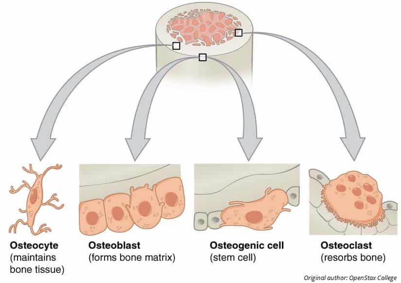
These diagrams clearly explain the functioning of the microscopes along with their respective parts. Download scientific diagram | structure of compact bone. Nov diagram for.net is a fully managed, extensible and powerful diagramming framework, which can help you create feature rich diagramming solutions in winforms, wpf, silverlight, xamarin.mac, monomac and asp. Usually bones that are thin and curved. Magnification by a microscope is the product of the individual magnifying ability of the oculars and the objectives. Sclerostin inhibits bone formation mostly by antagonizing lrp5/6, thus inhibiting wnt signaling. 3 mature bone cells, osteocytes, are found in tiny cavities within the matrix called lacunae. 8/30/2019 a) your histology atlas should include a labeled diagram of compact bone hyaline cartilage adipose blood b) each connective tissue will. This is a short tutorial using blender 2.8 that shows. These are mostly compacted bone with little marrow and include most of the bones in the limbs. The basic units of compact bone are called osteons or haversian systems. To better understand the structure and function of a microscope, we need to take a look at the labeled microscope diagrams of the compound and electron microscope. Scarica histology of human compact bone tissue under microscope view for education.
To better understand the structure and function of a microscope, we need to take a look at the labeled microscope diagrams of the compound and electron microscope. Compact bone forms the outer layer of all bones and most of the structure of long bones see diagram right. Under the microscope dense, compact bone shows a definite and a characteristic pattern of arrangement. Diagram of a compound microscope. 8/30/2019 a) your histology atlas should include a labeled diagram of compact bone hyaline cartilage adipose blood b) each connective tissue will.
33 2c Connective Tissues Bone Adipose And Blood Biology Libretexts from s3-us-west-2.amazonaws.com Des milliers de nouvelles images de grande qualité ajoutées chaque jour. Electron microscope image of trabecular bone (x100 magnification). These are mostly compacted bone with little marrow and include most of the bones in the limbs. Scarica histology of human compact bone tissue under microscope view for education. Compact bone, also called cortical bone, is the hard, stiff, smooth, thin, white bone tissue that surrounds all bones in the human body. Compact bone diagram under microscope. Bone tissue cross section diagram human oasissolutions co. These diagrams clearly explain the functioning of the microscopes along with their respective parts.
This human bone section shows the haversian canal (or osteon) structure of compact bone tissue. Download scientific diagram | structure of compact bone. The compact bone is composed of calcified extracellular material, the bone matrix and 3 major cell types which are * osteoblast which ssynthesize and secrete the organic for nerves, refer to snell's book of clinical anatomy. The transmitted brightfield digital images above were recorded using a qx3 microscope that was modified for auxiliary illumination. Label compact and spongy bone illustrations as demonstrated in class. Des milliers de nouvelles images de grande qualité ajoutées chaque jour. 3 mature bone cells, osteocytes, are found in tiny cavities within the matrix called lacunae. Sclerostin inhibits bone formation mostly by antagonizing lrp5/6, thus inhibiting wnt signaling. Between the rings of matrix, the bone cells (osteocytes) are located in spaces called lacunae. Compact bone microscope slide labeled learn by taking a quiz; If you look at compact bone under the microscope, you will observe a highly organized arrangement of concentric circles that look like tree trunks. Compact bone diagram bone cross section diagram file624 diagram of compact bone new. They consist of two outer layers of compact bone and an inner layer.
Decalcified compact bone at 60x magnification. Fruit microscopic structure of long bone. This is a short tutorial using blender 2.8 that shows. The transmitted brightfield digital images above were recorded using a qx3 microscope that was modified for auxiliary illumination. Online quiz to learn compact bone microscope slide labeled ;
Bone Structure And Function from 2012books.lardbucket.org Compact bone, also called cortical bone, is the hard, stiff, smooth, thin, white bone tissue that surrounds all bones in the human body. Sclerostin inhibits bone formation mostly by antagonizing lrp5/6, thus inhibiting wnt signaling. Compact bone diagram under microscope. 8/30/2019 a) your histology atlas should include a labeled diagram of compact bone hyaline cartilage adipose blood b) each connective tissue will. To better understand the structure and function of a microscope, we need to take a look at the labeled microscope diagrams of the compound and electron microscope. The larger ovals are blood vessels running through the bone. Each group of concentric circles (each tree) makes up the microscopic structural unit of compact bone called an osteon (this is also called a haversian. The inner surface of compact bone is lined by a thin, cellular layer, the endosteum.
For example if the ocular is 10x, and objective is 40x, the specimen is magnified 400 times.
The ground substance of bone is arranged in concentrated layers (lamellae) round the small canals which run parallel to the long axis (shaft) of the bone. Compact bone, spongy if you looked at it through a microscope, however, you would see that it's actually filled with many very. The compound microscope is more complicated than just a microscope with more than one lens. Bone basics and bone anatomyhave you ever seen fossil remains of dinosaur and ancient human each bone in your body is made up of three main types of bone material: Each group of concentric circles (each tree) makes up the microscopic structural unit of compact bone called an osteon (this is also called a haversian. Online quiz to learn compact bone microscope slide labeled ; Sclerostin inhibits bone formation mostly by antagonizing lrp5/6, thus inhibiting wnt signaling. Trouvez des images de stock de compact bone micrograph light microscopy bone en hd et des millions d'autres photos, illustrations et images vectorielles de stock libres de droits dans la collection shutterstock. Microscopic osteology and bone formation. Usually bones that are thin and curved. (micrograph provided by the regents of university of michigan. Having been constructed in the 16th century, microscopes have revolutionalized science with their ability to magnify small objects such as microbial cells, producing images with definitive structures that are. 2 compact bone we know that compact bone is very dense it is also very complex when viewed under a microscope.
Spongy bone and compact bone compact bone diagram. Confocal microscopy of fibroblast cells.


0 Comments